

Diagnostic imaging
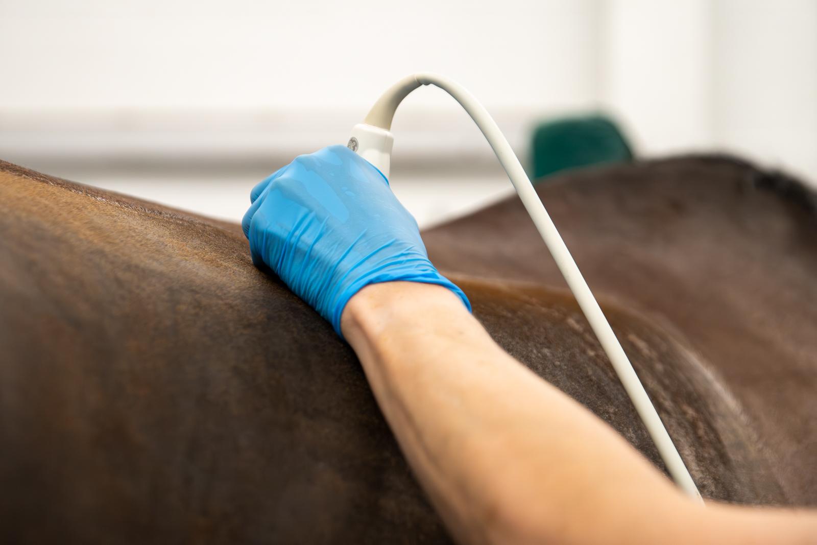
MRI, CT, Nuclear scintigraphy (bone scan), Radiography, Ultrasound and our Standing CT
Magnetic Resonance Imaging (MRI)
MRI is an excellent tool for evaluating the structures within the foot, and we also have experience of using it for image structures up to and including the carpus and tarsus.
MRI scans are performed by our qualified equine veterinary nurses, who also have training in equine diagnostic imaging and can obtain the best quality images possible.
The use of MRI has revolutionised our approach to foot lameness and penetrating injuries of the foot with a definitive diagnosis being achieved in 90% of cases. The use of MRI can save huge amounts of time and money that can be wasted in the absence of a definitive diagnosis, and then allows for specific treatments to be undertaken. MRI should be considered early in cases of foot lameness that cannot be diagnosed by traditional methods.
MRI scans are performed standing under sedation and do not require a general anaesthetic. Further information on MRI and its uses in equine medicine is available at www.hallmarq.net. Our referral team, led by Jonathon Dixon BVetMed MVetMed DipECVDI MRCVS are always happy to discuss cases that you think may benefit from an MRI scan and can be contacted on imaging@rainbowequinehospital.co.uk for further advice.